Skip to main menu Skip to main content Skip to footer

Glaucoma

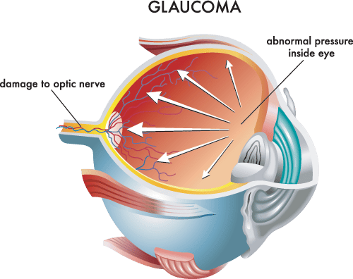
Glaucoma, often called the ‘Silent Thief of Sight,’ can strike without warning. It is a progressive condition in which the major nerve of the eye, the optic nerve, is damaged due to increased internal eye pressure.

As many as 65 million people around the world have glaucoma and it is the leading cause of blindness and visual impairment in the United States today. It is estimated that over four million people have glaucoma in the U.S. but only half of them know it. The key to combating this silent thief of sight is early detection followed by ongoing management of the disease.

What is Glaucoma?

Glaucoma consists of a group of conditions which damage the optic nerve. This nerve is responsible for taking light from the retina and sending impulses to the brain which are perceived as vision. Damage to the optic nerve dramatically diminishes eyesight.

At Wheaton Eye Clinic, glaucoma doctors perform an initial comprehensive glaucoma evaluation to:

  • measure the pressure in your eye (tonometry)
  • inspect your eye’s drainage angle (gonioscopy)
  • inspect your optic nerve and photograph it (ophthalmoscopy)
  • test your side, or peripheral, vision (visual field test)
  • measure the thickness of your cornea (pachymetry)
  • evaluate the thickness of the optic nerve fibers with nerve fiber analysis (eg. OCT)

Symptoms

The two most common types of glaucoma present themselves very differently. There are few or no symptoms or warnings for Primary Open-Angle Glaucoma (POAG). On the other hand, people with Angle-Closure Glaucoma (ACG) sometimes experience gradual loss of peripheral vision, usually in both eyes, which then advances to tunnel vision. Other people experience more pronounced symptoms including severe eye pain and headache, sometimes accompanied by nausea and vomiting, as well as blurred vision, halos around lights, sudden visual disturbances in low light and reddening of the eye.
Causes/Risk Factors

The highest risk factors for glaucoma are elevated internal eye pressure, which can only be detected during a doctor’s examination, and age. People are six times more likely to get glaucoma if they are over 60 years old.

In addition, glaucoma is the leading cause of blindness among African Americans who are six to eight times more likely to get glaucoma than Caucasians. African Americans should begin to have their eye pressure monitored before age 30. Hispanic populations and people of Asian descent also face an increased risk of glaucoma.

Family history and individual medical conditions present other risk factors. Current medical findings indicate glaucoma may have a genetic link that causes members of some families to be unusually susceptible to the disease.

Medical conditions such as diabetes, high blood pressure, heart disease or even nearsightedness can increase a person’s risk for glaucoma. Using corticosteroid medications for prolonged periods of time, especially in eye drop form, also appears to create risk of glaucoma. Major eye injuries can cause glaucoma to occur immediately after the injury or even years later.

Treatment Options

Depending on the type of glaucoma being experienced, your doctor may suggest treatment involving medication and/or surgery to lower the pressure in the eye and prevent further damage to the optic nerve.

If you are prescribed a medication for your glaucoma, it is important to carefully follow your medication regimen in order to maintain a healthy eye pressure and prevent vision loss. Your doctor may prescribe a combination of medications, or change your prescription over time, to reduce side effects or provide more effective treatment.

Sometimes, when medication is not producing the desired reduction in eye pressure, your doctor may suggest surgery. Glaucoma surgery involves either laser treatment or filtering microsurgery to reduce fluid build-up in the eye which, in turn, reduces intraocular pressure. Your doctor will recommend the best surgical treatment depending on the type and severity of your glaucoma and the general health of your eye. Surgery can help lower eye pressure when medication is not sufficient, however surgery cannot reverse vision loss.

Laser surgery is often recommended before filtering microsurgery unless eye pressure is very high or the optic nerve is badly damaged. During laser surgery, a tiny but powerful beam of light is used to make several small scars in the eye’s trabecular meshwork (the eye’s drainage system). The scars help increase the flow of fluid out of the eye which lowers the intraocular pressure.

When laser surgery does not successfully lower eye pressure, or the pressure begins to rise again, the doctor may recommend filtering microsurgery. This surgical procedure, called a trabeculectomy or sclerostomy, involves creating a tiny drainage hole in the sclera (the white part of the eye) with a small surgical tool.

In most cases, there is no pain involved during either laser treatment or microsurgery because local anesthetic and relaxing medications are used. Both are outpatient procedures. Within a few days after surgery, you will have a follow up appointment with your eye doctor to check eye pressure and look for any signs of infection or increased inflammation

In general, patients can resume normal daily activities the next day after laser surgery. For at least one week after filtering microsurgery, patients are advised to keep water out of the eye and avoid driving, reading, bending and doing any heavy lifting.

Following both laser and filtering microsurgery, medications often continue to be necessary in order to control and maintain eye pressure. Surgery may, however, lessen the amount of medication needed. Although there is a small risk of developing cataracts after some types of surgery for glaucoma, the potential benefits of surgery usually outweigh any risks.

If medication and surgery fail to lower intraocular pressure, your doctor may suggest an implant as a way to enhance standard glaucoma surgery. By positioning a device to keep the surgically-created drainage opening from healing and closing down, the surgeon is able to help decrease eye pressure by increasing outflow of fluid from your eye. Many current implants include a tube through which the aqueous fluid passes. Other implants are solid and promote the flow of fluid along the surface of the implant. Regardless of the type, all implants have the same goal—to increase outflow of fluid and decrease intraocular pressure.

Several thousand implants are used in the United States each year. Most of these are performed on what may be considered the more “complicated” glaucomas including cases of congenital glaucoma, glaucoma associated with diabetes or glaucoma following injury to the eye. In implant surgery, most of the device is positioned on the outside of the eye (toward the back) under the conjunctiva. Regardless of the particular implant used, fluid drains into the back end of the implant and is naturally reabsorbed by blood vessels. Although the implant is a foreign body, and there can be an inflammatory reaction immediately following surgery, stabilization usually occurs within 4–6 weeks.

Want to Know More?

Wheaton Eye Clinic is here to help.
We believe excellent patient care must also be convenient. Need to schedule an appointment? Choose from any of our locations.

Glaucoma Doctors

Locations

Wheaton Eye Clinic’s unparalleled commitment to excellence is evident in our continued growth. Today we provide world-class medical and surgical care to patients in six suburban locations—Wheaton, Naperville, Hinsdale, Plainfield, St. Charles, and Bartlett.

(630) 668-8250 (800) 637-1054
WARNING: Internet Explorer does not support modern web standards. This site may not function correctly on this browser and is best viewed on Chrome, Firefox or Edge browsers. Learn More.