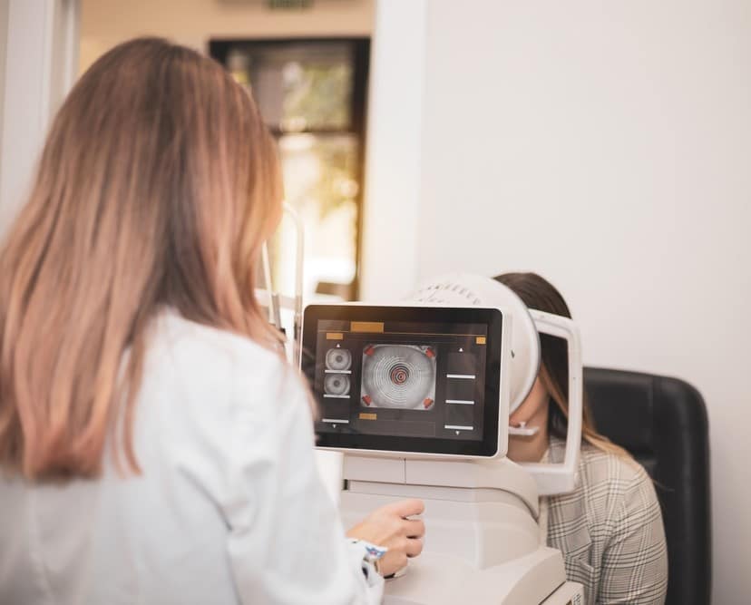
Dystrophies are inherited conditions that usually develop in both eyes. Depending on the dystrophy, it can present earlier or later in life. Usually, dystrophies are slowly progressive; some decrease vision and some do not. Different dystrophies affect different layers of the cornea and exhibit a variety of symptoms. There are many types of dystrophies but the most common are:
This is the most common dystrophy of the cornea. Many cases are asymptomatic, but some patients may experience distorted/blurry vision, while others may experience recurrent spontaneous painful corneal erosions.
Epithelial basement membrane dystrophy causes irregular thickening of the superficial outer layer of the cornea. Rather than being smooth and even, the surface develops thickened areas that cause wavy lines, dots, or cysts. It is typically bilateral, though symptoms may not be. This distortion to the outer layer of the cornea can sometimes result in blurry vision, ghosting, or recurrent corneal erosions.
A variety of treatment options include:
This is a dominantly inherited dystrophy that causes white “granular” looking spots to accumulate in the cornea, with clear spaces between the deposits. The granules are made up of a substance called hyaline.
Over time, vision can gradually decline due to the density of these granular deposits in the cornea. Some patients can also experience painful recurrent corneal erosions.
When vision is significantly decreased, or painful recurrent corneal erosions occur, a laser PTK (phototherapeutic keratectomy) treatment can be performed, though sometimes a corneal transplant is indicated.
In this dominantly inherited condition, glass-like branching lines made of amyloid accumulate in the cornea.
Vision can gradually decline due to these deposits, and recurrent corneal erosions can also commonly occur.
Some patients can benefit from PTK laser treatments, though others may require a corneal transplant.
This is the least common of the stromal dystrophies and is recessively inherited. It typically occurs in the first decade of life, between age 5-9 years. Mucopolysaccharide deposits in the cornea, causing severe diffuse haze and decreased vision.
Due to severe haze that develops, these patients typically need cornea transplantation to regain vision.
Fuchs’ endothelial corneal dystrophy is quite common and has an estimated incidence of 4-7%. Fuchs’ dystrophy, is characterized by loss of the cells that comprise the deepest layer of the cornea, a single layer of cells called endothelial cells. Two problems occur as a consequence that will affect vision. Guttae develop in the central cornea, causing a dimpled or “beaten bronze” texture to the inside layer of the cornea, thus causing loss of visual quality. Later in the disease process, the cornea can experience swelling which causes further vision decline.
The early symptoms are loss of contrast sensitivity and gradual loss of vision quality. Once swelling occurs, vision can be noticeably blurrier in the morning, which can somewhat improve as the day progresses. At advanced stages of corneal swelling, blisters can develop on the surface of the eye, causing foreign body sensation and pain.
If swelling is present, hypertonic sodium chloride drops and/or ointment can be used to treat some of the swelling, but this can have a limited effect on vision quality. Corneal transplantation of the deepest layer of the cornea (endothelial keratoplasty) is required to remove the guttae, replace the diseased endothelial cells, and improve vision.
DMEK is a type of partial-thickness cornea transplant which replaces only the deepest layers of the cornea. The patient’s diseased layer of endothelium and its adjacent basement membrane, called Descemet’s membrane, are removed from the eye and replaced with human donor corneal tissue that provides healthy corneal endothelium without guttae. This outpatient surgery can re-establish excellent vision in patients with Fuchs’ dystrophy.
DSEK is another type of partial-thickness corneal transplant. Similar to DMEK, DSEK replaces diseased endothelium and guttae with a healthy layer of donor endothelium but is a slightly thicker donor tissue. DSEK can be performed in eyes that may not be a good candidate for DMEK for a variety of reasons and would be determined by the operating surgeon.
Wheaton Eye Clinic’s unparalleled commitment to excellence is evident in our continued growth. Today we provide world-class medical and surgical care to patients in six suburban locations—Wheaton, Naperville, Hinsdale, Plainfield, St. Charles, and Bartlett.
(630) 668-8250 (800) 637-1054